Monday, 27 February 2017

Clean Room Technology in ART Clinics: A Practical Guide


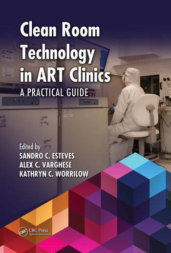
A new book of interest 'Clean Room Technology in ART Clinics: A Practical Guide', edited by Sandro C. Esteves, Alex C. Varghese, Kathryn C. Worrilow.

Regulatory agencies worldwide have issued directives or such requirements for air quality standards in embryology laboratories. This practical guide reviews the application of clean room technology or controlled environments specifically suited for Assisted Reproductive Technology (ART) Units. Its comprehensive coverage includes material on airborne particles and volatile organic compounds, including basic concepts, regulation, construction, materials, certification, clinical results in humans, and more.

Tim Sandle has contributed a chapter, titled 'Clean room design principles: Focus on particulates and microbials'.

Further details about the book can be found here.

Sandle, T. (2017) Clean room design principles: Focus on particulates and microbials. In Esteves, S. C., Varghese, A. C., and Worrilow, K. C. (Eds.) Clean Room Technology in ART Clinics: A Practical Guide, CRC Press, Boca Raton, U.S., pp75-91

Posted by Dr. Tim Sandle

Monday, 20 February 2017

The European approach to disinfectant qualification


Tim Sandle has written an overview of the European approach to disinfectant testing for the A3P publication La Vague.

Here is the introduction:

Contamination control is of great importance to healthcare facilities and to pharmaceutical cleanrooms. One way of ensuring the hygiene is maintained is through a cleaning and disinfection regime. After a disinfectant has been chosen based on its chemical properties and expected performance/effectiveness, each disinfectant should be validated to ensure its efficacy. Efficacy is demonstrated through performance testing to show that the disinfectant is capable of reducing the microbial bioburden in either suspension (planktonic state) or from cleanroom surfaces to an acceptable level.

The article can be read here.


Posted by Dr. Tim Sandle


Thursday, 16 February 2017

New target for taming Ebola


A team of scientists led by Ronald Harty, a professor of pathobiology and microbiology at the University of Pennsylvania's School of Veterinary Medicine, has identified a mechanism that appears to represent one way that host cells have evolved to outsmart infection by Ebola and other viruses. In a new paper, he and colleagues reveal that host cells sequester viral proteins away from the plasma membrane within the cell, thus preventing viruses from spreading.

Harty says that finding a way to amplify this molecular interaction could lead to a novel antiviral strategy against Ebola that could temper the pathogen's damage.

The scientists knew that, in infections with Ebola as well as many other viruses, including Marburg, rabies and HIV, viral matrix proteins, such as Ebola VP40, interacted with host proteins through short protein motifs: the PY motif on the viral protein and the WW motif on the host cell protein. Prior to this current study, all of the known interactions enabled the virus to bud efficiently from the cell.

In the current work, the researchers screened for new WW motifs from mammalian cell proteins that bound tightly to the PY motif of Ebola virus' VP40 protein. Not every PY motif binds to every WW domain; the interaction is specific, like a lock and key.

The screen turned up some proteins that the Penn team had explored before but also a new one, a protein called BAG3, known as a chaperone protein, which under normal physiological conditions acts to promote cell survival.

After confirming that Ebola VP40 interacted with full-length BAG3 in mammalian cells specifically via the WW domain, the researchers went on to test its functionality in influencing budding. They used a test that avoids manipulating the actual Ebola virus, which is too dangerous for the Penn Vet laboratory. Instead, they examined virus-like particles, which are produced by the virus's matrix protein, VP40, and are not infectious but accurately mimic the budding step of infection.

When the researchers examined cells expressing either Ebola or Marburg VP40 and then added in BAG3, they found that VLP budding went down in a dose-dependent manner. When they mutated the WW domain of BAG3 so it couldn't interact with VP40's PY domain, budding levels remained unchanged.

Knocking down levels of naturally occurring BAG3 with synthetic strands of RNA did just the opposite, increasing budding.

"With all of these assays, there was a consistent effect on budding levels, either up or down," Harty said.

Enhancing such an interaction, perhaps in combination with other therapies that attack the virus at other stages of its life cycle, could give the immune system the opening it needs to overcome an infection.

Using confocal microscopy and fluorescently labeled proteins, the scientists discovered that BAG3 appeared to be sequestering VP40 in the cell's cytoplasm away from the plasma membrane where the viral particles would need to go in order to bud off and spread to infect other cells. Their work identified BAG3 as the first WW containing host protein to negatively affect virus budding.

Although the group has not yet tested the VP40-BAG3 interaction with live Ebola or Marburg virus -- those experiments are planned -- they did find that BAG3 limits budding of a recombinant vesicular stomatitis virus that contains the Ebola PY motif.


"We used that to show that this works in a live virus infection," Harty said. "Taken together, we're hoping and assuming that it works the same way with the authentic Ebola virus."

In addition to testing BAG3 interactions with the Ebola and Marburg virus, Harty's lab also plans to further investigate what BAG3 is doing to VP40, whether it's simply sequestering it or whether it's modifying or degrading it.

See:

Jingjing Liang, Cari A. Sagum, Mark T. Bedford, Sachdev S. Sidhu, Marius Sudol, Ziying Han, Ronald N. Harty.Chaperone-Mediated Autophagy Protein BAG3 Negatively Regulates Ebola and Marburg VP40-Mediated Egress. PLOS Pathogens, 2017; 13 (1): e1006132 DOI:10.1371/journal.ppat.1006132

Posted by Dr. Tim Sandle

Wednesday, 15 February 2017

Good Practice Guidelines for Blood Establishments


EU directive integrates Council of Europe Good Practice Guidelines for blood establishments

Good Practice Guidelines have been prepared through co-operation between the European Directorate for the Quality of Medicines & HealthCare of the Council of Europe (EDQM) and the Commission of the European Union (EU). The elaboration of the Good Practice Guidelines included substantial public consultations giving stakeholders an opportunity to comment on the draft version.

The Good Practice Guidelines were adopted by the European Committee (Partial Agreement) on Blood Transfusion (CD-P-TS) of the Council of Europe during its plenary session in November 2016. The document is an integral part of the 19th Edition of the Council of Europe Guide to the Preparation, Use and Quality Assurance of Blood Components, Appendix to Recommendation No. R (95) 15 of the Committee of Ministers, referred to below as the “Guide”.

Following adoption of the Good Practice Guidelines by the European Commission, European Union and European Economic Area Member States shall ensure that Blood Establishments take fully into account the standards and specifications set out in those Guidelines when implementing their quality system, in line with the new Commission Directive (EU) 2016/1214. In order to bring into force the laws, regulations and administrative provisions necessary to comply with this Directive by 15 February 2018, some member states need to translate the text of the Good Practice Guidelines into their national language. To expedite this process, a verbatim copy of the text of the Good Practice Guidelines that will be published in the Guide is downloadable as a document here.

Posted by Dr. Tim Sandle

Sunday, 12 February 2017

Antimicrobial resistant bacteria detected in air pollution


Areas subject to air pollution contain a growing concentration of bacteria that are resistant to antimicrobials, according to a new study. Consequently, air movement may provide a new ways for their transmission.
By Tim Sandle

The study, highlighting a growth in airborne or air-carried antimicrobial resistant bacteria, has come from China, with a focus on Beijing. The extent of antimicrobial resistance has been assessed at the genetic level, with the detection of DNA from genes that make bacteria resistant.
The study has been led by Professor Joakim Larsson, of the Sahlgrenska Academy and the University of Gothenburg. Professor Larsson is especially concerned about air as a potential vector for spreading antimicrobial resistant pathogens.
Antimicrobial resistance (including resistance to antibiotics) is the biggest risk faced by human societies. The implications are that life expectancy could fall due to people dying from diseases that are readily treatable today.
With the new research, Professor Larsson’s team tested air samples for genes that make bacteria resistant to antibiotics. From a total of 864 samples, DNA was tested and related back to its source. One area of concern was the detection of a series of genes that provide resistance to carbapenems. These are a group of ‘last resort antibiotics’ and they are administered for infections caused by bacteria that the most resistant to antimicrobials. Carbapenems are members of the beta lactam class of antibiotics, which kill bacteria by binding to penicillin-binding proteins and inhibiting cell wall synthesis.
Importantly, the findings cannot indicate if the bacteria are alive in the air. This narrows the risk somewhat; however, the potential for genetic transfer with viable bacteria, upon contact, remains.
The study is, however, only a starting point. The researchers plan to assess whether antimicrobial resistance can indeed spread through air. Here the focus will move to a study of the air within the vicinity of European sewage treatment plants. Sewage plants are regularly called out as potential sites for the spread of antimicrobial resistance. This is because, with so many different types of bacteria coming together in sewage plants, this provided optimal conditions for organisms to swap genes that confer resistance. This also means antibiotic-resistant bacteria could evolve much faster than they would in isolation.
Data will be cross-checked in relation to the air and to the sewage. Additional samples will be taken from people who live in close proximity to sewage plants.
The research findings are published in the journal Microbiome and the research paper is titled “The structure and diversity of human, animal and environmental resistomes.”

Thursday, 9 February 2017

New surgical mask renders viruses harmless


Hyo-Jick Choi, a professor in the University of Alberta Department of Chemical and Materials Engineering, noticed that many people wear a simple surgical-style mask for protection during outbreaks of influenza or other potentially deadly viruses such as severe acute respiratory syndrome (SARS) or Middle East respiratory syndrome (MERS).

Trouble is, the masks weren't designed to prevent the spread of viruses.

"Surgical masks were originally designed to protect the wearer from infectious droplets in clinical settings, but it doesn't help much to prevent the spread of respiratory diseases such as SARS or MERS or influenza," says Choi.

Airborne pathogens like influenza are transmitted in aerosol droplets when we cough or sneeze. The masks may well trap the virus-laden droplets but the virus is still infectious on the mask. Merely handling the mask opens up new avenues for infection. Even respirators designed to protect individuals from viral aerosols have the same shortcoming -- viruses trapped in respirators still pose risks for infection and transmission.

Masks capable of killing viruses would save lives, especially in an epidemic or pandemic situation. During the 2014-2015 season nearly 8,000 Canadians were hospitalized with the flu. That same year, deaths related to influenza in Canada reached an all-time high of nearly 600.

Knowing that the masks are inexpensive and commonly used, Choi and his research team went about exploring ways to improve the mask's filter. And this is where a problem he is struggling with in one field of research -- the development of oral vaccines like a pill or a lozenge -- became a solution in another area.

A major hurdle in the development of oral vaccines is that when liquid solutions dry, crystals form and destroy the virus used in vaccines, rendering the treatment useless. In a nifty bit of engineering judo, Choi flipped the problem on its head and turned crystallization into a bug buster, using it as a tool to kill active viruses.

Choi and his team developed a salt formulation and applied it to the filters, in the hope that salt crystals would "deactivate" the influenza virus.

The mechanics of simple chemistry make the treatment work. When an aerosol droplet carrying the influenza virus contacts the treated filter, the droplet absorbs salt on the filter. The virus is exposed to continually increasing concentrations of salt. As the droplet evaporates, the virus suffers fatal physical damage when the salt returns to its crystalized state.

While developing solid vaccines, Choi observed that sugar used for stabilizing the vaccine during the drying process crystalizes as it dries out. When crystals form, sharp edges and spikes take shape and they physically destroy the virus vaccine.

"We realized that we could use that to our advantage to improve surgical masks," said Choi.

In a series of experiments and tests at the University of Alberta and in the Department of Medical Zoology at the Kyung Hee University School of Medicine in Seoul, South Korea, the team arrived at a perfect treatment that improves the efficacy of the fibre filter inside the masks.

By using a safe substance (table salt) to improve an existing, approved product, Choi sees very few roadblocks to implementing the innovation.

For further details see:

Fu-Shi Quan, Ilaria Rubino, Su-Hwa Lee, Brendan Koch, Hyo-Jick Choi. Universal and reusable virus deactivation system for respiratory protection. Scientific Reports, 2017; 7: 39956 DOI: 10.1038/srep39956


Posted by Dr. Tim Sandle

Monday, 6 February 2017

Disinfectants: Properties, Applications and Effectiveness


A new book of interest has been published,m titled "Disinfectants: Properties, Applications and Effectiveness." The book is edited by Ana Sofia Cardoso, Cristina Maria Martins Almeida, Telma Costa Cordeiro, and Vanessa de Jesus Gaffney. The book is published by Nova, New York.

Antiseptics and disinfectants are extensively used at home, in occupied buildings, recreational areas, industries (the water industry, food processing industry and pharmaceutical industry, among others), hospitals and other healthcare settings for a variety of topical and hard-surface applications. They play a critical role in controlling the spread of environmentally transmitted pathogens in healthcare and food-processing environments, as well as at home. A wide variety of active chemical agents are found in these products, many of which have been used for hundreds of years for antisepsis, disinfection, and preservation.

Although its main purpose is to control human exposure to microorganisms through preventive action, its use should also be carefully controlled in order to prevent healthcare problems that may consequently emerge due to their toxicity.

The problems regarding the use of disinfectants are not new, although unquestionably tangible and pertinent, due to its broad application in the referred economical activities, as well as due to the development and emerging of new compounds with this activity.

This book aims to address the various scenarios regarding the use of disinfectants. Accordingly, through its eleven chapters it is possible to become aware of the wide range of disinfectant applications, as well as the concerning advantages and limitations of its use. This book is divided into two main sections. The first section, after an overview regarding the use of disinfectants in society, addresses questions related to its toxicology and health repercussions along with microbiological mechanisms.

In the second section, a far-reaching exploration of the application of disinfectants in a set of specifically selected economic activities, alongside issues concerning their environmental impact and regulatory matters is addressed. This section also includes two case studies on novel disinfection methods.

Tim Sandle has contributed a chapter on disinfectants in the pharmaceutical sector. The reference is:

Sandle, T. (2016) Disinfectants in the Pharmaceutical Industry. In Cardoso, A. S., Almeida, C. M. M., Cordeiro, T. C. and  Gaffney, V.J.  (Eds.) Disinfectants: Properties, Applications and Effectiveness, Nova Science Publishers, New York, pp109-142

The book is available via Amazon:


Posted by Dr. Tim Sandle

Sunday, 5 February 2017

Innovative Strategies for Reducing Nasal Colonization


Staphylococcus aureus (S. aureus) and methicillin resistant S. aureus (MRSA) can cause difficult-to-treat infections due to their antibiotic resistance. Approximately 90,000 people are affected yearly, and 20,000 people die from infections caused by bacteria like MRSA. Research shows that nasal antiseptics are a highly effective means of reducing nasal colonization of bacteria and subsequent infection.

Methicillin-resistant Staphylococcus aureus (MRSA) is a bacterium responsible for several difficult-to-treat infections in humans. MRSA is any strain of Staphylococcus aureus that has developed, through horizontal gene transfer and natural selection, multi- resistance to beta-lactam antibiotics, which include the penicillins (methicillin, dicloxacillin, nafcillin, oxacillin, etc.) and the cephalosporins. MRSA evolved from horizontal gene transfer of the mecA gene to at least five distinct S. aureus lineages. Strains unable to resist these antibiotics are classified as methicillin-susceptible Staphylococcus aureus, or MSSA. The evolution of such resistance does not cause the organism to be more intrinsically virulent than strains of S. aureus that have no antibiotic resistance, but resistance does make MRSA infection more difficult to treat with standard types of antibiotics and thus more dangerous.

This following paper details the impacts of hospital acquired infections financially and medically, and provides proven strategies on achieving nasal decolonization.

Posted by Dr. Tim Sandle

Saturday, 4 February 2017

Captive monkeys gain human microbiomes


To explore how a microbiome adapts to a new environment, University of Minnesota researcher Dan Knights and his colleagues turned their attention to nonhuman primates.

According to Bioscience Techniques, the team examined the microbiota from two species of monkeys living in a zoo, in a sanctuary, or in the wild using shotgun sequencing. The data showed that monkeys living in the wild possessed broad, diverse microbial signatures, while the animals living in sanctuaries showed considerably less variety. Interestingly, monkeys living in zoos showed even less diversity, with their microbes actually resembling signatures found in modern humans. A follow-up study of 33 monkeys representing 8 different species raised in a zoo showed a similar microbial complement.

For further details see:

Clayton JB, Vangay P, Huang H, Ward T, Hillmann BM, Al-Ghalith GA, Travis DA, Long HT, Tuan BV, Minh VV, Cabana F, Nadler T, Toddes B, Murphy T, Glander KE, Johnson TJ, Knights D. Captivity humanizes the primate microbiome. Proc Natl Acad Sci U S A. 2016 Sep 13;113(37):10376-81.

Posted by Dr. Tim Sandle

Friday, 3 February 2017

Scientists tissue-engineer functional part of human stomach


Scientists have used pluripotent stem cells to generate human stomach tissues in a Petri dish that produce acid and digestive enzymes. They grew tissues from the stomach's corpus/fundus region. The study comes two years after the same team generated the stomach's hormone-producing region (the antrum). The discovery means investigators now can grow both parts of the human stomach to study disease.

The discovery means investigators now can grow both parts of the human stomach to study disease, model new treatments and understand human development and health in ways never before possible.

See:

Kyle W. McCracken, Eitaro Aihara, Baptiste Martin, Calyn M. Crawford, Taylor Broda, Julie Treguier, Xinghao Zhang, John M. Shannon, Marshall H. Montrose, James M. Wells. Wnt/β-catenin promotes gastric fundus specification in mice and humans. Nature, 2017; DOI: 10.1038/nature21021

Posted by Dr. Tim Sandle

Special offers