Saturday 29 April 2017

10 Easy Steps To Clean Spills In A Biosafety Cabinet


For over 40 years, NuAire has been providing laboratory equipment that better enables researchers to work under defined environmental conditions. A biosafety cabinet or biological safety cabinet (BSC) is an enclosed, ventilated laboratory workspace for users to safely handle materials that might contain pathogens. There are several different models of BSCs, which are differentiated by the user’s experimental focus and the degree of bio-containment required.

The primary purpose of a BSC is to protect the laboratory worker and the surrounding environment from pathogens such as bacteria and viruses being used within the cabinet. All exhaust air is filtered through HEPA-filters as it exits the biosafety cabinet, removing the harmful pathogens. Most classes of BSCs have a secondary purpose that is to maintain the sterility of materials inside the cabinet.

It happens at some point to even the most seasoned laboratory user that a spill occurs within the BSC. Taking precautionary measures before and during your work with hazardous materials could help keep you and others safe. Remember, if a spill occurs, don’t panic.  Here are some simple steps to keep you and your laboratory safe. Please check with your EHS office or Biosafety Officer to ensure your have proper steps in place in case of a spill based of standard Biosafety in Microbiological and Biomedical Laboratories (BMBL).

Spill Kit

The lab should have a kit or the components readily available to address an accidental spill. This includes an easy-to-read outline of the spill response Standard Operating Procedures (SOPs) that should be posted, read and understood by everyone in the lab, the appropriate personal protection equipment (PPE); including eye protection, a clean lab coat or scrubs and spare slip-on shoes in case clothing contamination occurs. In addition, absorbent materials, disinfectant (e.g., 10% bleach), tongs or forceps to pick up broken containers and a biohazard waste container are needed.

Wear appropriate personal protection equipment (PPE)

Before beginning your work in the BSC, be sure to dress appropriately wearing the approved PPE designated for your laboratory.

At a minimum, laboratory coats should be worn buttoned over street clothing, protective eyewear should be on at all times and latex or nitrile gloves are necessary when handling culture, contaminated surfaces, or equipment.

Again, be sure to follow the recommended BMBL procedures for the biosafety level of the laboratory you are working in.

Perform decontamination steps while the cabinet is operating

When a spill of bio-hazardous material occurs within a BSC, cleanup should begin immediately, while the cabinet continues to operate. Keeping the cabinet running will prevent the escape of airborne contaminants and ensure that whatever is in the cabinet stays in the cabinet protecting those around you and the laboratory.

Remove items from the spill area

Before attacking the spill, first remove the tubes, pipettes or any other item that might have contained the spilt liquid and place them into the biohazard bag in the cabinet. It is important to contain contaminated materials inside the operating cabinet to avoid exposure to the laboratory. Always use tongs or forceps to pick up any glass or sharps to prevent accidental injury.

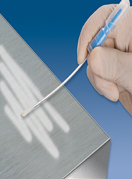
Cover the spill with absorbent material

Cover the spill inside the BSC with absorbent material such as paper towels and let the spill soak in. This helps to prevent aerosolization of the contaminant. Once the towel is covering the spill, apply appropriate disinfectant for the type of spill onto the towel, working from the outer edge to the middle of the towel. Applying the disinfectant from the outside to the inside of the spill helps to trap the material within the paper towel and decontaminant. It is important to note that the agent spilled must not be resistant to the disinfectant selected for cleanup. Having a laboratory procedure that addresses the biohazards you might encounter will ensure that you have the appropriate materials available for a spill. Bleach solutions have several advantages over the others such as low cost, fast acting and broad spectrum of effectiveness, but they are corrosive for use on stainless steel surfaces inside a BSC and should be rinsed (refer to step 7).

Allow 20 minutes for disinfectant contact time

Depending on what material was spilled and what disinfectant you are using, you might need to vary the disinfectant reaction time. As a rule of thumb, 20 minutes should be adequate time to neutralize the contaminant.

Wipe up spill and excess liquids with towels

Once the spill has been contained and the disinfectant has had adequate time to react, use the towels to wipe up excess liquid. Place used towels into a biohazard bag located in the cabinet.

Treat the area with the decontaminant again

Apply disinfectant to the spill area again and give it appropriate time to work before wiping up with fresh towels. This helps ensure that all of the contaminated material and surface are decontaminated. Also check the spill pan under the work surface and disinfect following the same procedure if needed.

Rinse the spill area well


If bleach (or any other corrosive disinfecting agent) was used to clean the spill, use sterile water to rinse and then again to wipe the residual bleach (or disinfectant) off of the working surface. Bleach is very corrosive to stainless steel and will cause damage over time if it is used to clean the cabinet.

Once the cabinet has been cleaned, remove gloves and other protective equipment

Thoroughly wash your hands with soap and water. Run the BSC for at least 10 minutes before resuming work. Report the spill incident to your supervisor.

Following these steps will help you keep yourself and those around you safe if a spill in the BSC occurs. It will also help to maintain your equipment for years of use. So keep the workspace clean and let the research flow!

For more information please visit www.nuaire.com or call 1.800.328.3352


Original source: Pharmaceutical Online

Thursday 27 April 2017

Tracking yellow fever virus replication


Yellow fever virus (YFV) is a member of the flavivirus family that also includes Dengue and Zika virus. The virus, which is thought to infect a variety of cell types in the body, causes up to 200,000 cases of yellow fever every year, despite the widespread use of a highly effective vaccine. The vaccine consists of a live, attenuated form of the virus called YFV-17D, whose RNA genome is more than 99 percent identical to the virulent strain. This one percent difference in the attenuated virus' genome may subtly alter interactions with the host immune system so that it induces a protective immune response without causing disease.

To explore how viruses interact with their hosts, and how these processes lead to virulence and disease, Alexander Ploss, assistant professor of molecular biology, and colleagues at Princeton University adapted a technique -- called RNA Prime flow -- that can detect RNA molecules within individual cells. They used the technique to track the presence of replicating viral particles in various immune cells circulating in the blood of infected mice. Mice are usually resistant to YFV, but Ploss and colleagues found that even the attenuated YFV-17D strain was lethal if the transcription factor STAT1, part of the antiviral interferon signaling pathway, was removed from mouse immune cells. The finding suggests that interferon signaling within immune cells protects mice from YFV, and that species-specific differences in this pathway allow the virus to replicate in humans and certain other primates but not mice.

Accordingly, YFV-17D was able to replicate efficiently in mice whose immune systems had been replaced with human immune cells capable of activating interferon signaling. However, just like humans immunized with the attenuated YFV vaccine, these "humanized" mice didn't develop disease symptoms when infected with YFV-17D, allowing Ploss and colleagues to study how the attenuated virus interacts with the human immune system. Using their viral RNA flow technique, the researchers determined that the virus can replicate inside certain human immune cell types, including B lymphocytes and natural killer cells, in which the virus has not been detected previously. The researchers found that the panel of human cell types targeted by the virus changes over the course of infection in both the blood and the spleen of the animals, highlighting the distinct dynamics of YFV-17D replication in the human immune system.


The next step, said Florian Douam, a postdoctoral research associate in the Department of Molecular Biology and first author on the study, is to confirm YFV replication in these subsets of immune cells in YFV-infected patients and in recipients of the YFV-17D vaccine. Viral RNA flow now provides the means to perform such analyses.

To find out more see:

Florian Douam, Gabriela Hrebikova, Yentli E. Soto Albrecht, Julie Sellau, Yael Sharon, Qiang Ding, Alexander Ploss. Single-cell tracking of flavivirus RNA uncovers species-specific interactions with the immune system dictating disease outcome. Nature Communications, 2017; 8: 14781 DOI: 10.1038/NCOMMS14781

Posted by Dr. Tim Sandle

Tuesday 25 April 2017

Changes in gut microbiota after unhealthy diet examined


The intestine is covered with a plethora of microorganisms, collectively termed gut microbiota, that are thought to play an important role in regulating the metabolism and shaping the immune system. Many studies have shown that dysbiotic bacteria can cause disease. However, these studies generally follow a similar protocol that may impact on the outcome: They transfer dysbiotic bacteria to axenic mice that do not have any microbiota. For example, axenic mice that receive microbiota from the gut of obese mice would increase their total body fat, indicating that microbiota play a causative role in the development of obesity.

Researchers have taken a different approach to addressing the role of microbiota. The researchers reasoned that axenic mice are ill-equipped to deal with dysbiotic microbiota. Their gut barrier is impaired, favoring an uncontrolled spread of bacteria throughout the body. In addition, their immune system is not well developed. Thus, instead of using axenic mice as recipients of dysbiotic microbiota, the team used normal, healthy mice, which have not been treated with antibiotics before. In contrast to previous research, the team found that a dysbiosis is not necessarily harmful. In fact, it may even lead to metabolic adaptions that protect the body against disease.
High fat diet increases the production of glucose by the liver and can eventually lead to metabolic disease. However, when researchers transplanted dysbiotic microbiota from mice on high-fat diet to healthy mice, they found that the production of glucose in the liver was reduced rather than increased. Therefore, dysbiotic microbiota counter the metabolic effect of high-fat diet and may thus protect the host from its consequences. Similar effects were also observed when the researchers used microbiota from genetically obese mice.

In another set of experiments, the researchers transplanted dysbiotic microbiota from obese mice to healthy mice and then put these mice on a high-fat diet. Normally, a high-fat diet would lead to weight gain. However, the body mass of mice that had received dysbiotic microbiota did not change, and their adipose tissue showed smaller fat cells, consistent with increased plasma free-fatty acids.

In conclusion, dysbiosis after high-fat diet may not all be detrimental. As long as the gut barrier is intact and the immune system is functional, dysbiosis may even protect the body from metabolic effects of unhealthy diets.

See:


Simon Nicolas, Vincent BlascoBaque, Audren Fournel, Jerome Gilleron, Pascale Klopp, Aurelie Waget, Franck Ceppo, Alysson Marlin, Roshan Padmanabhan, Jason S Iacovoni, François Tercé, Patrice D Cani, JeanFrançois Tanti, Remy Burcelin, Claude Knauf, Mireille Cormont, Matteo Serino. Transfer of dysbiotic gut microbiota has beneficial effects on host liver metabolism. Molecular Systems Biology, 2017; 13 (3): 921 DOI: 10.15252/msb.20167356


Posted by Dr. Tim Sandle

Sunday 23 April 2017

Medical technologists find cheaper way to make essential medicine


Medical technologists have found a means to create an anti-fungal medication, designed to combat Cryptococcal meningitis, less expensively. The drug is intended for use in parts of Africa.
A fungal form of meningitis causes a major problem in parts of Africa, and it can lead to in-excess of 600,000 deaths each year. The aggressive disease accounts for close to 20 percent of deaths associated with AIDS related Human Immunodeficiency Virus (HIV) infections globally, based on U.S. Centers for Disease Control and Prevention figures.
To combat incidences of the fungal infection, medics have profiled that an existing medicine could help. However, the medication is prohibitively expensive for health systems in many parts of Africa. As a solution, medical researchers have come up with a low-cost way of manufacturing the drug. This should lead to greater use of the drug in those parts of the world that need it the most.
The drug in question is the anti-fungal drug flucytosine. The drug has been used in countries like the U.S. for several decades. The World Health Organization, in 2011, made the recommendation that patients with Cryptococcal meningitis, an infection associated with those infected with HIV take flucytosine (in combination with another medication called amphotericin B) as a first line of defense. the primary agent of infection is Cryptococcus neoformans.
To make the medication at a lower cost, PharmPro reports that Dr. Graham Sandford from Durham University (U.K.) has come up with a new method. This is make flucytosine out of readily available, naturally occurring cytosine. This is by pumping inexpensive fluorine gas and a solution of cytosine in formic acid through a steel tube, where flucytosine is produced by later recrystallization.
The development of the alternative medication is described in the American Chemical Society journal Organic Process Research & Development. The research paper is titled "One-Step Continuous Flow Synthesis of Antifungal WHO Essential Medicine Flucytosine Using Fluorine."


Purchase:


Posted by Dr. Tim Sandle

Saturday 22 April 2017

Teixobactin: A Powerful Tool for Combating Resistant Strains


Resistance to antibiotics has grown out to be serious health dilemma. Despite this serious health crisis, no new antibiotics have been revealed since last 30 y. A new ray of hope in the form of teixobactin has come out of the dark which can prove to be effective in defeating resistance. This new antibiotic has an interesting mechanism of action against bacteria. The discovery of this wonderful compound has evolved as major breakthrough especially in this era of antibiotic catastrophe. This review article highlights various facets of teixobactin. Its chemistry, mode of action, in vitro and in vivo aspects have been thrown light upon in this review. Though the compound has not undergone clinical trial studies but its effect in mice models has given a hope for overpowering resistance. This article has been mainly communicated with an objective to provide information about teixobactin which has emerged as ray of hope for fighting antibiotic resistance.

This relates to a new paper of interest, see: IJPS

Wednesday 19 April 2017

Faster, more accurate detection of food- and water-borne bacteria


Recently, Charles S. Henry and colleagues developed a paper-based method to detect Salmonella, Listeria and E. coli in food and water samples. In their latest study, Henry's team wanted to see if it would be feasible to use this paper-based technique in conjunction with electrochemical analysis to produce more refined results.


To simulate contaminated food, the researchers exposed clean alfalfa sprouts to E.coli and Enterococcus faecalis bacteria. They also collected unfiltered water from a nearby lagoon. For colorimetric detection, the team built a simple light box, which served as a substitute for a laboratory plate reader. Then they used a smartphone to take a series of images of the 84 paper-based well plates over time. For the electrochemical portion of the experiment, they used a series of electrodes printed onto plastic transparency sheets. Both approaches used the same assays to successfully detect harmful bacteria in the samples within 4 to 12 hours, and both produced complementary findings. They conclude that combining their paper-based technique with electrochemistry could lead to a simpler, yet more comprehensive way to detect bacterial contaminants in food and water.

See:

Jaclyn A. Adkins, Katherine Boehle, Colin Friend, Briana Chamberlain, Bledar Bisha, Charles S. Henry. Colorimetric and Electrochemical Bacteria Detection Using Printed Paper- and Transparency-Based Analytic Devices. Analytical Chemistry, 2017; DOI: 10.1021/acs.analchem.6b05009

Posted by Dr. Tim Sandle

Monday 17 April 2017

Understanding the Importance of Safety in Pharmaceutical Manufacturing and Transportation


Pharmaceutical manufacturing and transportation is an industry that requires a high level of oversight to make sure the medications being manufactured meet the required specifications and that they are being transported correctly to ensure maximum efficacy upon delivery. For many patients, these medications can do everything from improve the quality of their life to ensure their survival, which is what makes these regulations so important.

Special guest post by Megan Ray Nichols

What do you need to understand about safety in pharmaceutical manufacturing and transportation?

Manufacturing

The first step in pharmaceutical safety happens in the manufacturing stage. In the United States, manufacturers are constantly under scrutiny by the FDA (Food and Drug Administration) to ensure each batch of a medication meets the same quality and efficacy standards as the previous batches.
Pharmaceutical companies that produce their products in the United States are subject to the FDA’s Current Good Manufacturing Practice, or CGMP. These practices ensure the company is using the highest-quality raw products as well as up-to-date manufacturing technology to provide a standard level of quality across all of their products.

This is essential, especially for over-the-counter medications, because most customers don’t have the skills or equipment necessary to test their medication and make they’re safe and effective. For a bottle of Tylenol, for example, most people don’t look closely enough to see anything other than they’ve got the proper number of pills in hand. It’s not laziness on the consumer’s part, but rather a sign of the trust they’ve placed in the manufacturer.

Transportation

Safety plays a significant role in the transportation of medications. Once the medication is complete and safe to transport, the problem becomes a logistical one. A number of different variables have to be taken into account, including:

·         Form: In what form are these drugs being transported? Solid — in the form of pills or powder — or liquid, in IV bags or sterile bottles? Are they gaseous substances that need to be transported in pressurized containers?

·         Type: Security for the shipment of controlled substances should be much higher because they are most likely to be stolen and sold on the street.

·         Requirements: Do the medications have to be kept at a certain temperature to maintain safety and efficacy?

·         Destination: Where are the medications going? Will there be multiple transport changes (such as truck to plane) for final delivery, or will the products remain in the same vehicle for their entire journey?
These variables and many more have to be taken into account when planning the best way to produce and deliver pharmaceutical products. Items that have to be kept at a low temperature, for example, will need to be more carefully monitored than those that can be stored at room temperature.
Security at loading and unloading points is also a necessity — especially for controlled substances at risk for theft.

Customer Expectations

Customer expectations are higher than they’ve ever been, in part because they know they can put their trust in these companies and that there are laws in place to ensure every Tylenol or Excedrin they take is going to be close to identical. That trust comes from experience — they know that if there is a problem, the product will be removed until it can be fixed, like the cross-contamination problem that Novartis experienced in 2012 with Excedrin and four of its other over-the-counter products.

The applicable safety regulations are the key to making sure the trust of these consumers is not misplaced.

Regulations concerning the safety of pharmaceutical manufacturing and transportation aren’t there just to pacify bureaucrats and legislators — they’re in place to ensure that safe and effective medication is available to patients around the world. By ensuring this safety throughout both the manufacturing and shipping processes, we can help to ensure that no matter their destination, all of these medications arrive where they’re needed most in time to help.

Microbial swab recovery


Swabs are commonly used as part of microbiological environmental monitoring programmes and it is important to understand the suitability of the swabs used and their limitations.

A new paper by Ravikrishna Satyada and Tim Sandle looks into this issue. The abstract for the paper reads:

Surface monitoring by using swabs forms a regular part of environmental monitoring of cleanrooms. There are different factors that affect swab recovery, from tip type to enumeration method. One factor, for swabs where the microorganisms are detached from the swab tip and which are then membrane filtered, is the period of vortex mixing. This paper discusses microbial surface sampling, and the factors that affect swab recovery. The paper presents some experimental data where vortex times are considered for a range of microorganisms. The study outcome indicates that 15 seconds vortex mixing is sufficient to obtain microbial recoveries from the swab tip above 50%.

The reference is:

Satyada, R. and Sandle, T. (2016) Releasing capacity of pre-sterile cotton swabs for discharging sampled microorganisms, European Journal of Parenteral and Pharmaceutical Sciences, 21 (4): 121-128

For further details please contact Tim Sandle

Best buy:


Posted by Dr. Tim Sandle

Sunday 16 April 2017

'Smart' bacteria remodel their genes to infect our intestines


Infectious diarrhea, a common disease of children, is responsible for over 2 million infant deaths annually in developing counties alone. A primary cause of this and other devastating conditions is enteropathogenic bacteria, which attack the intestinal tract when contaminated food is consumed.

The infection process involves hundreds of genes and proteins, both in the infectious bacteria and the human host. However, the processes by which the pathogens establish themselves in our gut are poorly understood.

Now, a new study published in the journal Science, by researchers at the Hebrew University of Jerusalem's Faculty of Medicine, describes how pathogens sense their host, and tailor their gene expression to exploit their host to cause disease. The research was led by led by Prof. Ilan Rosenshine, the Etta Rosensohn Professor of Bacteriology at the Hebrew University.


Working with a pathogenic strain of E. coli, the researchers found that the bacteria can sense attachment to the human intestinal cells and activate gene expression in response. This was demonstrated by engineering one of these genes to express a protein that stains the expressing bacteria to appear green under the microscope. Under microscopic examination, the researchers observed that only the attached bacteria fluoresce in bright green, whereas non-attached bacteria remain dark.

The researchers also deciphered how upon sensing that it has attached to intestinal cells, the pathogen reorganizes its gene expression, including genes involved in virulence and metabolism, to exploit the host cell. These findings may lead to the development of new strategies to combat bacterial infection.

See:

Naama Katsowich, Netanel Elbaz, Ritesh Ranjan Pal, Erez Mills, Simi Kobi, Tamar Kahan, Ilan Rosenshine. Host cell attachment elicits posttranscriptional regulation in infecting enteropathogenic bacteria. Science, 2017; 355 (6326): 735 DOI: 10.1126/science.aah4886


Posted by Dr. Tim Sandle

Thursday 13 April 2017

3-D tissue culture models to mimic human gut infections


Central to the development of tissue models that can better predict how humans respond to infection is the understanding that cells and tissues in our bodies function in a three-dimensional (3-D) context. Accordingly, cell-based models of tissues made in the laboratory must be developed with the same appreciation for the 3-D tissue microenvironment encountered by pathogens in the body.

While this research concept has long been appreciated by the cancer and regenerative medicine world, the infectious disease world has been slower to get on board.

Now, an ASU Biodesign Institute team has developed and applied 3-D tissue models to study bacterial infectious disease nearly two decades ago -- and spearheaded the adoption of 3-D tissue models as a new paradigm to study infectious disease -- has reported their latest advancement in 3-D intestinal model development.

The new study, a collaboration between Arizona State University and NASA's Johnson Space Center, was led by Cheryl Nickerson, a researcher at ASU's Biodesign Institute and professor in the School of Life Sciences.

Their united goal is to develop more realistic models of intestinal tissue to thwart Salmonella, a leading cause of food poisoning and systemic disease worldwide with many varieties causing severe and sometimes fatal infections with an economic impact in the billions of dollars.

Interestingly, the response of this new model to infection with the different types of Salmonella was very different for each strain, thus demonstrating the model's ability to distinguish between these closely related pathogens based on their infection characteristics. Specifically, important differences were observed between the bacterial strains in model colonization (adherence, invasion and intracellular survival) and intracellular co-localization patterns in epithelial and immune cells.

See:

Jennifer Barrila, Jiseon Yang, Aurélie Crabbé, Shameema F. Sarker, Yulong Liu, C. Mark Ott, Mayra A. Nelman-Gonzalez, Simon J. Clemett, Seth D. Nydam, Rebecca J. Forsyth, Richard R. Davis, Brian E. Crucian, Heather Quiriarte, Kenneth L. Roland, Karen Brenneman, Clarence Sams, Christine Loscher, Cheryl A. Nickerson. Three-dimensional organotypic co-culture model of intestinal epithelial cells and macrophages to study Salmonella enterica colonization patterns. npj Microgravity, 2017; 3 (1) DOI: 10.1038/s41526-017-0011-2


Posted by Dr. Tim Sandle

Monday 10 April 2017

Biodecontamination of Cleanrooms and Laboratories Using Gassing Systems


Tim Sandle has written a new article on the decontamination of cleanrooms and laboratories by gassing systems:

Cleanrooms, laboratory areas, isolators and biosafety workspaces (microbiological safety cabinets) require a level of cleanliness and microbial control (achieved through disinfection) according to the intended use of the area.

This paper assesses the current technologies and process steps required for the effective biodecontamination of cleanrooms and containment laboratories.

The reference is:

Sandle, T. (2017) Biodecontamination of Cleanrooms and Laboratories Using Gassing Systems, Journal of GxP Compliance, 21 (1): 1-12

For details, see: IVT or contact Tim Sandle

Best buy:


Posted by Dr. Tim Sandle

Friday 7 April 2017

Phage therapy shown to kill drug-resistant superbug


Phages are viruses that kill bacteria but are otherwise harmless. A major advantage is that phages only target the harmful bacteria, so there are less side of the effects often associated with antibiotics. Phage therapy however has not had the same level of funding as drug development, due to a lack of convincing pre-clinical efficacy studies.

Researchers have shown that phage therapy is highly effective in treating established and recalcitrant chronic respiratory tract infections caused by multi-drug resistant Pseudomonas aeruginosa strains. They show that phages are capable of killing the bacteria in long term infected lungs, such as those suffered by patients with the inherited disease Cystic Fibrosis, indicating a potential new therapeutic option for these hard to treat life threatening infections.

For further information see:

Elaine M Waters, Daniel R Neill, Basak Kaman, Jaspreet S Sahota, Martha R J Clokie, Craig Winstanley, Aras Kadioglu. Phage therapy is highly effective against chronic lung infections with Pseudomonas aeruginosa. Thorax, 2017; thoraxjnl-2016-209265 DOI: 10.1136/thoraxjnl-2016-209265


Posted by Dr. Tim Sandle

Monday 3 April 2017

Risk of microbial spores in cleanrooms


The risks posed by microbial spores pose to cleanrooms in terms of their survival mechanisms and, in the case of fungi, ease of dispersal, are substantial. One means to address this is through the use of a proven sporidical agent. This is the subject of a new article by Tim Sandle.

The article considers the use of sporicidal disinfectants, examining different types and considering the range of factors that affect sporicide efficacy. Importantly, the selection of sporicidal agents is not straightforward. Several types of sporicidal agent are extremely corrosive to stainless steel, plastic and soft metals as well as being a potential health hazard to operators. For this reason, such agents tend to be used sparingly alongside other disinfectants.

Reference:

Sandle, T. (2017) Risk of microbial spores to cleanrooms: Part 2: Selection of sporicidal disinfectants, Clean Air and Containment Review, Issue 29, pp14-16

For further details see: Spores

See also:


Posted by Dr. Tim Sandle

Special offers